
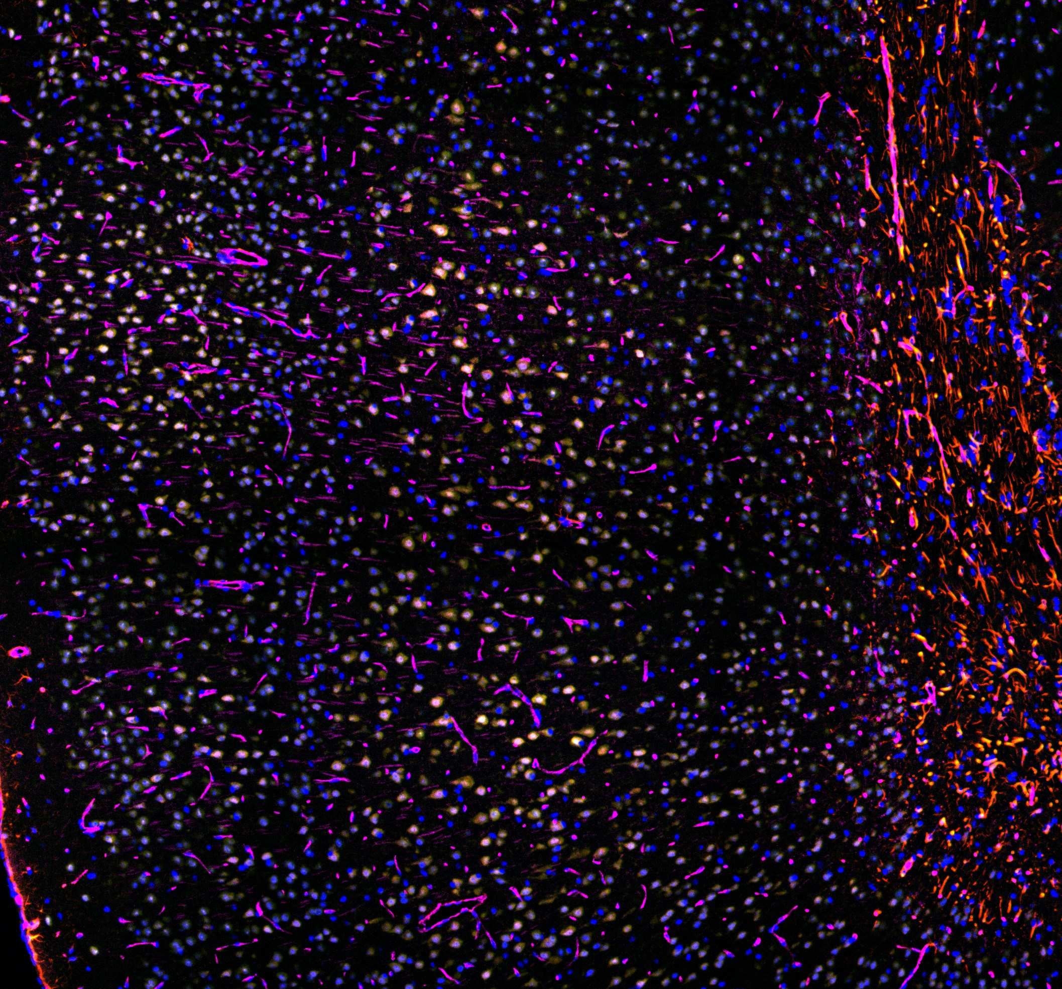
Immunofluorescence (IF)
Advanced Immunofluorescence (IF) services at Wax-it elevates your research through our single and multiplexing IF services. Our team of experts employs top-tier reagents and cutting-edge technologies in superior method development programs. Together, we ensure the precise localization and analysis of specific proteins within tissue samples, enriching your preclinical research endeavors with unparallel clarity and depth.
Applications of Immunofluorescence
Immunofluorescence at Wax-it offers a wide array of applications:
- Protein Localization: Visualizing the distribution of proteins within various cellular compartments such as the nucleus, cytoplasm, and mitochondria.
- Disease Diagnosis: Detecting specific antibodies in samples to aid in diagnosing infectious diseases and autoimmune disorders.
- Drug Discovery: Studying the uptake and effects of drugs marked with fluorescent probes.
- Immunophenotyping: Identifying cell surface antigens and facilitating studies on cell-cell interactions.
- Signal Pathway Analysis: Observing signaling molecules to understand cellular processes like proliferation and apoptosis.
- Cellular Structure Visualization: Aiding in the study of cellular dynamics through the visualization of microtubules, filaments, and organelles.
Types of Tissue Used in Immunofluorescence
Wax-it processes various tissue types for IF labeling:
- Cell Culture Samples: Analyzing protein expression in vitro.
- Fresh-frozen Tissue Sections: Visualizing protein distribution in snap-frozen samples.
- Formalin-fixed, Paraffin-embedded (FFPE) Tissue Sections: Examining protein expression in preserved tissues.
- Cultured Tissue Sections: Investigating proteins in a physiologically relevant setting.
Immunofluorescence Assays with Multiplexing
- Direct Immunofluorescence: Utilizes primary antibodies directly conjugated to fluorophores, minimizing background noise and speeding up the detection process.
- Indirect Immunofluorescence: Enhances sensitivity through the use of secondary antibodies, allowing for the detection of multiple antigens simultaneously.
- Double and Multiple Labeling: Identifies co-localized antigens using primary antibodies from different species.
- Multiplexing: Unlock the power of multiplexing with our innovative approach. Label your slides, strip, and relabel effortlessly to visualize a multitude of target proteins, maximizing insights and accelerating your research journey.
Customized Protocols and Advanced Capabilities
- Custom Protocols: Tailoring staining protocols for specific experiments, considering the biological specimen and target antigens.
- Validation: Following validated protocols with stringent quality control measures for reliable and reproducible results.
- Scalability and Multiplexing: Accommodating various project sizes and allowing simultaneous detection of multiple antigens in a single tissue sample.
- High-Quality Imaging: Employing advanced fluorescence microscopy, such as the Leica Thunder Imager and ZEISS Axio Scan 7 for detailed image analysis.
- Expert Consultation: Providing guidance throughout the staining process, to get the best outcome for the study.
- Efficiency: Ensuring swift turnaround times through the integration of expertise and technology.
- Quantification: Utilizing machine learning algorithms and sophisticated imaging software for accurate quantification of tissue images.
At Wax-it, our commitment to illuminating the path to discovery through cutting-edge IF solutions ensures unparalleled support for our clients’ research needs. Let us enhance your research with our comprehensive immunofluorescence services, providing detailed insights into complex biological processes.