
Customized Protocol Development
We specialize in developing precise, customized protocols tailored to the unique characteristics of each complex medical device. Considering factors such as material composition and implantation site, our protocols ensure accurate and reliable analysis. Through diligent collaboration with clients, we adhere to regulatory standards to drive robust research outcomes and informed decision-making in device development and assessment.

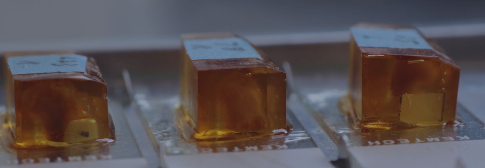
Resin Histology
Resin histology is a vital component in our medical device evaluations, providing detailed insights into tissue-device interactions at a microscopic level. The technique where we master cutting sections through the device with the tissue intact enables precise visualization of device components interacting with biological tissues, critical for assessing device performance and safety.

Cutting, Grinding and Polishing
Our histology team employs cutting-edge techniques to create 10- 20-micron sections, ensuring precise analysis of intricate structures and materials. By meticulously cutting, grinding, and polishing samples, we achieve optimal sectioning for detailed examination, overcoming complexities to provide accurate insights into device characteristics and their biological interactions.

Histochemical Staining
Utilizing a variety of histochemical staining and labeling techniques, we reveal insights into tissue and immune responses, yielding valuable information about device compatibility. A combination of staining (including H&E, Masson’s Trichrome, and Picrosirius Red and many other special stains) and IHC labeling provides insight into specific components such as collagen, elastin, and other connective tissues, helping us evaluate structural integrity and potential adverse effects. Moreover, this capability enables us to visualize inflammatory markers with precision - providing deeper insight into complex and rare cell types within the same tissue.

Imaging & Analysis
Our digital whole-slide scanning capabilities generate high-resolution images that vividly depict device-tissue interactions and alterations in tissue architecture at various magnifications. Wax-it encompasses facilities for standard as well as large-format slides. The production of thin sections through specialized resin-based histology allows us to achieve much higher resolution. In addition to whole-slide imaging, we also perform various quantifications, including area measurements, using high-resolution images.

Pathology Services
Our pathology services are supported by board-certified pathologists essential for regulatory compliance and scientific publication. Pathology scoring, displayed in data tables, facilitates rapid preliminary analysis, ensuring that our comprehensive reports meet the stringent standards required for regulatory submissions and scientific discourse.
At Wax-it, we are committed to unravelling the complexities of medical devices through histopathology, providing expert insights that shape the future of device development and assessment. By integrating state-of-the-art facilities, technological expertise, and rigorous quality assurance measures, we offer unparalleled service in medical device histopathology.
